
Reaching the eight-week mark of pregnancy can feel exciting and overwhelming all at once. Your baby is developing quickly, and many parents are eager to see what’s happening inside the womb for the first time.
Around this stage, your healthcare provider may recommend a first-trimester ultrasound to confirm that your pregnancy is progressing as expected. This early scan helps assess your baby’s growth, heartbeat, and location, while also ruling out potential concerns.
If you’re feeling nervous about what to expect, you’re not alone. Below, we’ll walk through what doctors are looking for at an 8-week ultrasound, how the exam works, and how to prepare so you can feel more confident going in.
What is an Ultrasound?
An ultrasound is a safe medical imaging test that uses high-frequency sound waves to produce images (also known as a sonogram) of the inside of your body, such as your internal organs and tissues.1 Two types of ultrasounds can be used during a prenatal appointment: transvaginal and transabdominal.
Transvaginal
Most commonly used early in pregnancy, transvaginal ultrasounds involve placing a wandlike device, called a transducer, inside the vagina. The transducer releases sound waves that create images of your cervix, fallopian tubes, ovaries, uterus, and pelvic region. This type of ultrasound can help confirm a pregnancy and monitor early development. It may also be used to detect your baby’s heartbeat and identify potential concerns.
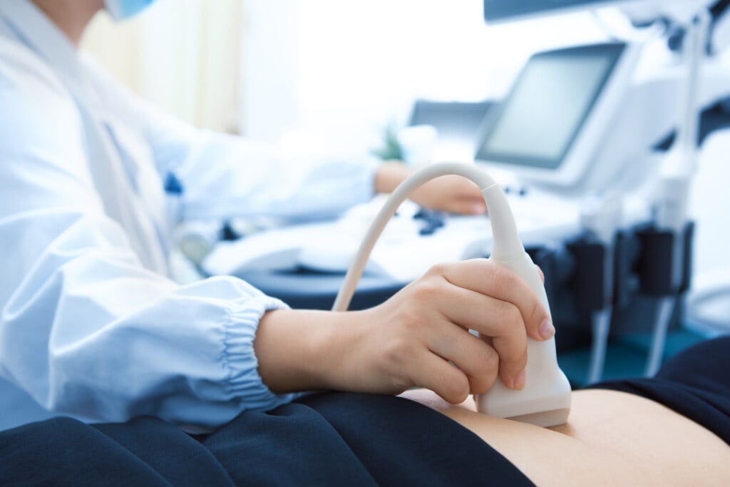
Transabdominal
Most people envision a transabdominal ultrasound when they hear the word “ultrasound.” It is a handheld device passed back and forth over a woman’s abdomen, covered in gel. During this process, the transducer emits sound waves across the belly and bounces them off the baby’s bones and tissues to create a picture of the baby. This allows expectant parents to see their baby on the screen.
Related: What Will My Baby Look Like? 7 Factors That Play a Role
Why Am I Getting an 8-Week Ultrasound?
An early ultrasound helps confirm important details about your pregnancy and ensures development is on track.
Not all pregnancies are the same, and not all women share the same experience. At this stage, a noninvasive ultrasound allows healthcare professionals to evaluate both your health and your baby’s development. It also helps your doctor monitor whether early growth is progressing as expected. Additional reasons include:
Gestational Age
Are you counting down the days until you can meet your newborn face-to-face? Your physician can give you a more accurate estimate of your baby’s due date during the ultrasound by measuring the gestational age of your pregnancy, or how far along you are.
Multiple Pregnancies
It’s all in the name with this one. Your physician will check how many fetuses are present in the uterus, with twins being the most common type of multiple pregnancy. Pregnancies involving three or more fetuses are rare.
Related: Signs You Might Be Having Twins or Multiples
Heartbeat
Nothing in the world will sound more magnificent than hearing your baby’s heartbeat for the very first time. During your ultrasound, you should be able to hear the beautiful thudding. A fetus’s heartbeat is typically detectable around six weeks of gestation and beats between 150 and 160 beats per minute.2
Ectopic Pregnancy
If you’re experiencing pelvic or abdominal pain or vaginal bleeding, your doctor will perform a pelvic ultrasound to rule out an ectopic pregnancy. This occurs when a fertilized egg has implanted outside the uterus, such as in the fallopian tubes, which is the most common location where it may occur. Other areas that can be affected include the cervix, cesarean scars, abdomen, or pelvis.
If you’re not feeling any pain, it’s still vital that your embryo’s location is confirmed. Although ectopic pregnancies are rare, occurring in about 1.4 percent of pregnancies, they can cause organ damage and are life-threatening if untreated.3
Related: Ectopic Pregnancy Symptoms To Look Out For
How Can I Prepare for an Ultrasound?
For a transvaginal ultrasound, minimal preparation is required. During your appointment, you’ll be asked to remove your clothing and any jewelry that can interfere with the test while in the examination room and be given a gown to put on. You’ll likely be asked to use the restroom to empty your bladder before the ultrasound.
On the other hand, your physician will request that you have a full bladder for the examination during a transabdominal ultrasound. You’ll need to consume about 32 ounces of water an hour before the test, since a full bladder helps lift the intestines to provide a clearer image of your organs. You can use the bathroom after your ultrasound if needed.
In comparison to the transvaginal test, you will not be required to wear a gown. Wear comfortable clothing that day, such as a loose shirt that you can easily lift to your chest when the cooling gel is applied to your belly.
What Happens After the Ultrasound?
If your physician performs the ultrasound, you may receive your results right away. When a technician conducts the exam, the images are sent to a radiologist for review and then shared with your healthcare provider. While the ultrasound itself typically takes about half an hour, final results may take up to 24 hours.
Afterward, your provider will explain what the findings mean for your pregnancy and answer any questions you may have. Although waiting can feel nerve-wracking, ultrasounds are safe and often provide valuable reassurance during early pregnancy. Take a moment to appreciate this milestone and the first glimpse of your growing baby.